Educational Resources
Embolization Products | Sample CaseWith recent advances in endovascular cerebral aneurysm repair, the accuracy of measuring aneurysm volume has become increasingly important. Calculating the percent packing volume after coil embolization can be used as an indicator of long term success and depends on an accurate calculation of aneurysm volume.1-2 Tamatani et al1 have shown a relationship between the percent packing volume and recanalization of aneurysms. They found that the incidence of recanalization was significantly lower in aneurysms packed greater than 25%. Sluzewski et al2 have shown a relationship between compaction of the coil mesh and percent packing volume. They reported no compaction at 6 months in aneurysms that were packed greater than 24%.
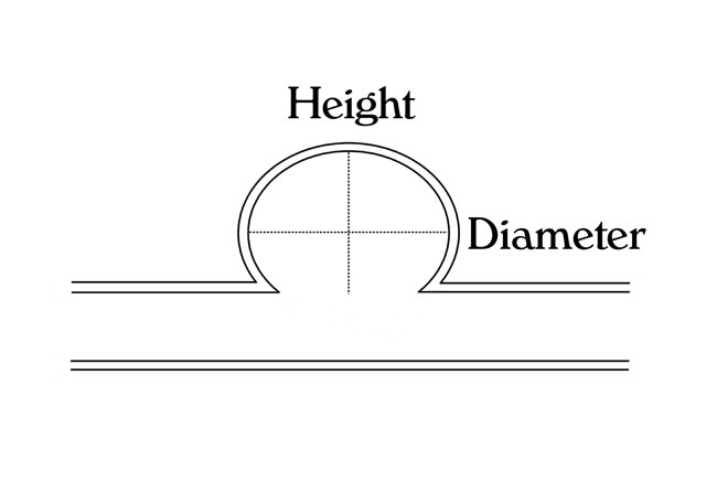
To calculate the percent packing volume, first calculate the volume of the aneurysm (Figure 1). The volume of the aneurysm can be obtained by using 3D volumetric software or manually by choosing a shape on the Home Page and entering the measured dimensions.
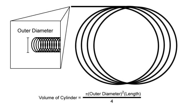
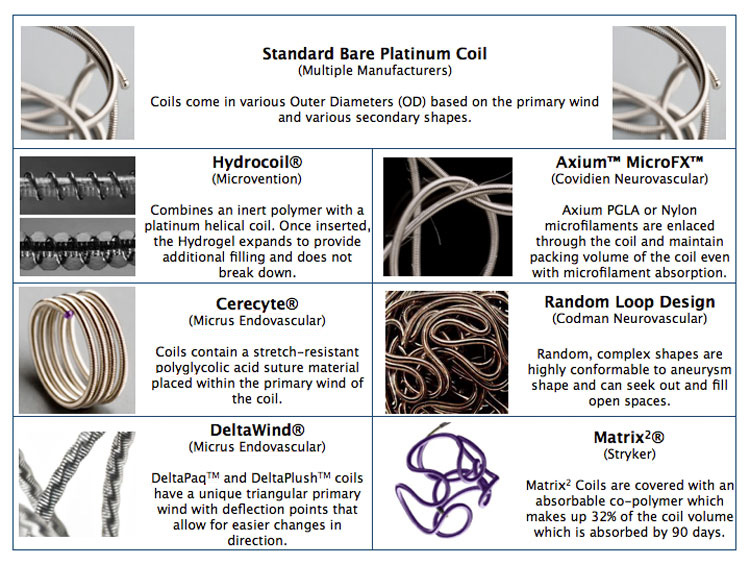
On the % Packing Page enter the total length of the coils deployed. Coil volume is determined using the formula for a cylinder. While most coils have a circular primary wind, there are slight variations which the Outer Diameters (OD) provided by the manufacturers reflects. The provided Outer Diameters (Figure 2) are listed on the % Packing Page. Finally, the percent packing volume is calculated by dividing the volume of the coils delivered by the volume of the aneurysm (Figure 3).
 |
 |
|
| Figure 1 - Aneurysm Volume | Figure 2 - Coil Volume | Figure 3 - % Packing Volume |
| (click thumbs to enlarge) | ||
Embolization Products
Sample Case
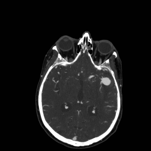
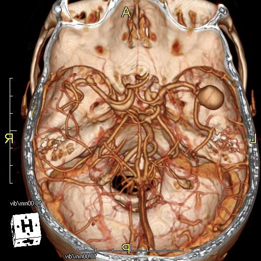
45 year old female presented with right arm weakness and a Head CT with and without contrast was performed. CT demonstrates subarachnoid hemorrhage with a left MCA aneurysm.
 |
 |
 |
| Image 1 - Non Contrast CT | Image 2 - CT Angiogram | Image 3 - CTA 3D Reformat |
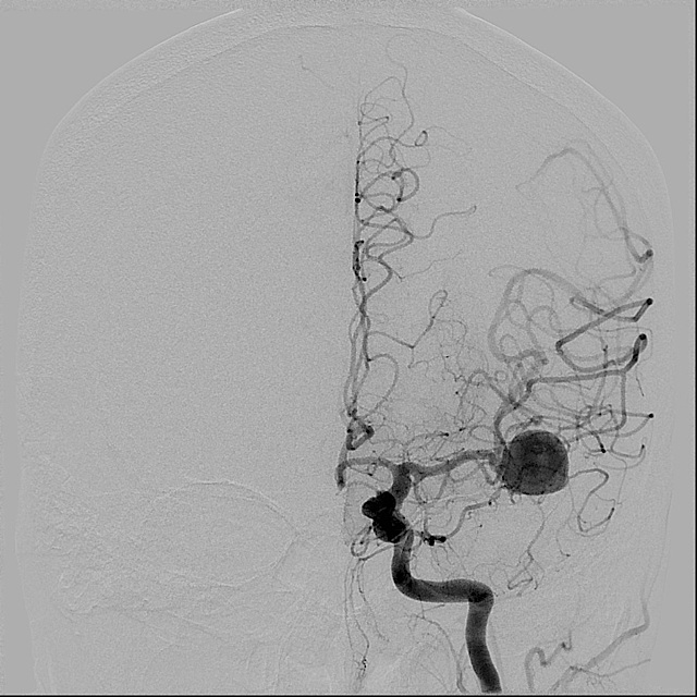
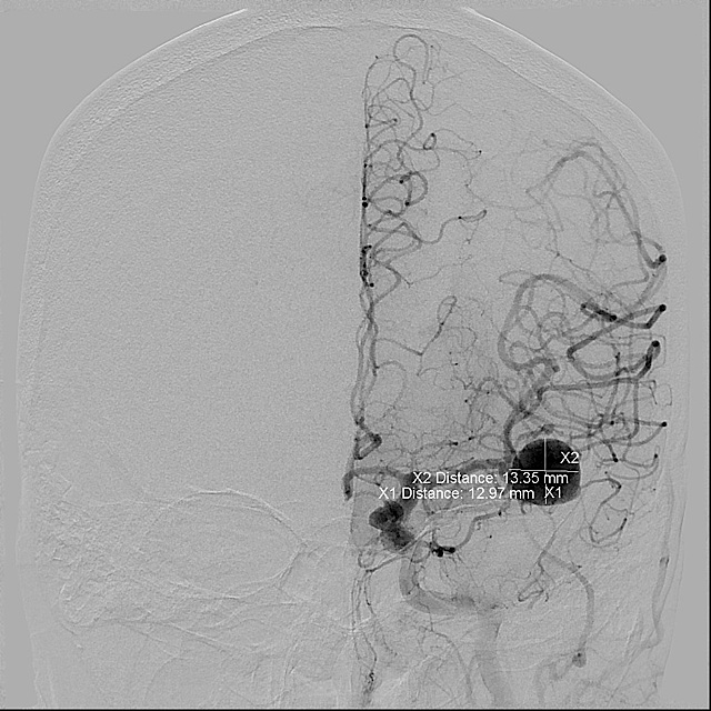
Cerebral angiography was then performed demonstrating the left MCA aneurysm. Aneurysm was measured in Image 6.
 |
 |
 |
| Image 4 - Cerebral Angiogram | Image 5 - Cerebral Angiogram | Image 6 - Measurements |
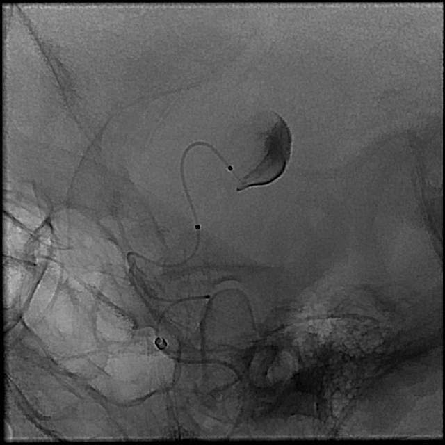
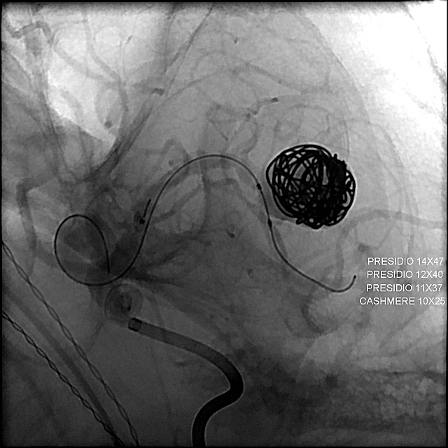
A microcatheter was advanced into the aneurysm and coils deployed (Images 7 and 8). Post procedure images demonstrate occlusion of the aneurysm (Image 9).
 |
 |
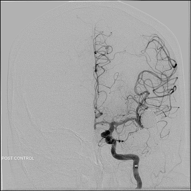 |
| Image 7 - Microcatheter | Image 8 - Coil Deployment | Image 9 - Final Result |
References
- Tamatani S, Ito Y, Abe H, et al: Evaluation of the stability of aneurysms after embolization using detachable coils: Correlation between stability of aneurysms and embolized volume of aneurysms. AJNR May 2002, 23:762-767.
- Sluzewski M, Willem JvR, Slob MJ, et al: Relation between aneurysm volume, packing, and compaction in 145 cerebral aneurysms treated with coils. Radiology 2004; 231:653-658.