
Sample Case
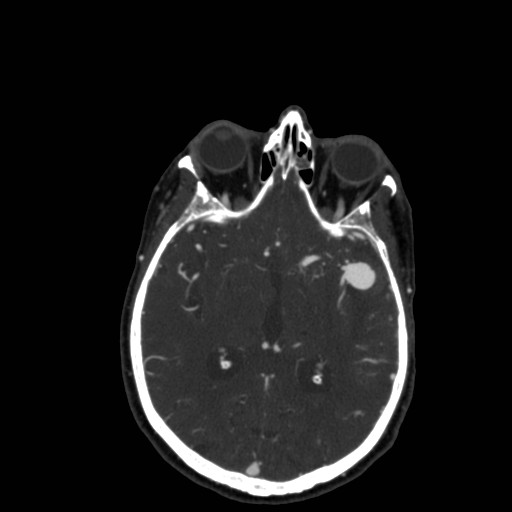
45 year old female presented with right arm weakness and a Head CT with and without contrast was performed. CT demonstrates subarachnoid hemorrhage with a left MCA aneurysm.
 |
 |
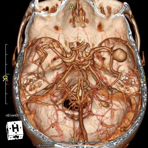 |
| Image 1 - Non Contrast CT | Image 2 - CT Angiogram | Image 3 - CTA 3D Reformat |
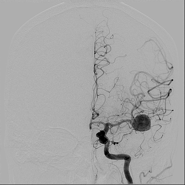
Cerebral angiography was then performed demonstrating the left MCA aneurysm. Aneurysm was measured in Image 6.
 |
 |
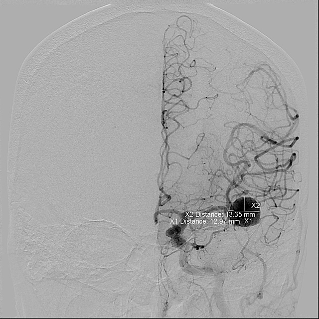 |
| Image 4 - Cerebral Angiogram | Image 5 - Cerebral Angiogram | Image 6 - Measurements |
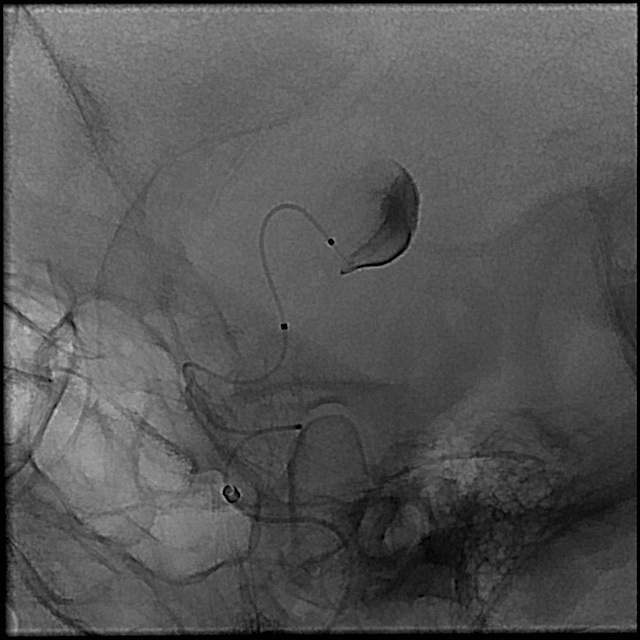
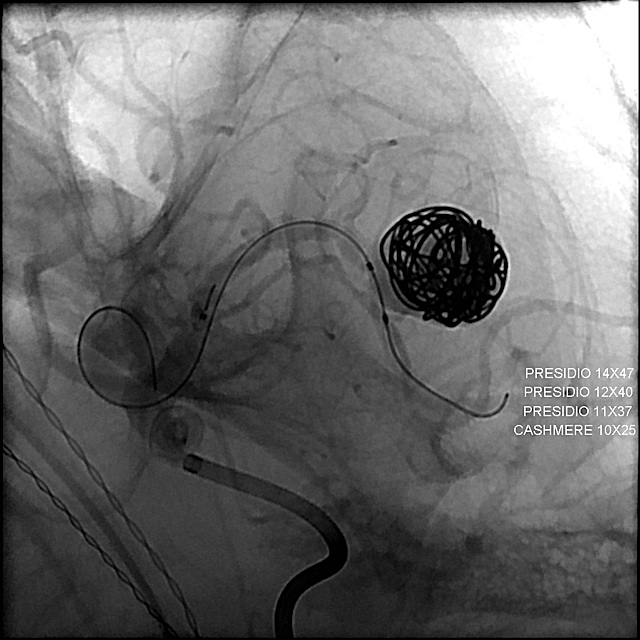
A microcatheter was advanced into the aneurysm and coils deployed (Images 7 and 8). Post procedure images demonstrate occlusion of the aneurysm (Image 9).
 |
 |
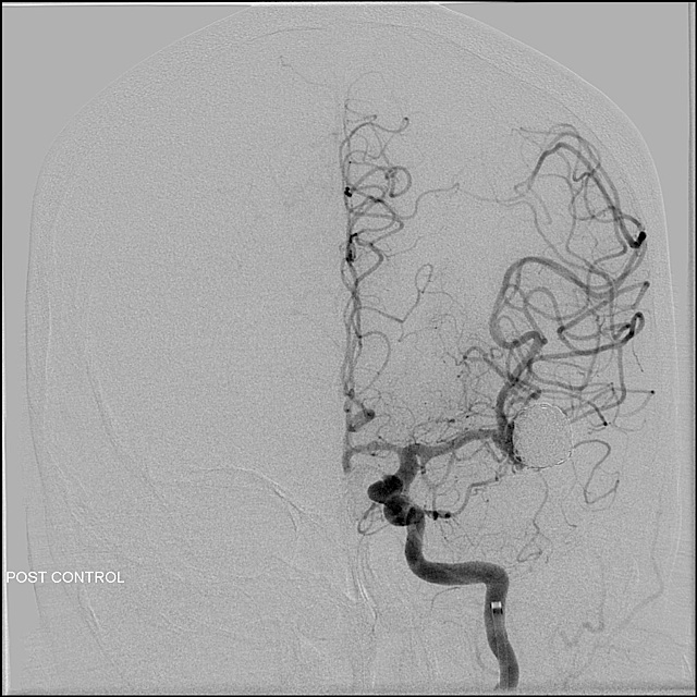 |
| Image 7 - Microcatheter | Image 8 - Coil Deployment | Image 9 - Final Result |